Key Points
Diabetic Retinopathy = Diabetic microangiopathy affecting the retinal blood vessels leading to progressive retinal damage and ultimately blindness
- Occurs due to poor metabolic control
- Often the first complication to develop
- Can lead to complete blindness
Epidemiology & Aetiology
- Diabetic retinopathy is the commonest cause of blindness in <65 year olds
- Risk factors:
- Poor metabolic control chronically
- A small change in Hba1c has a massive impact
- Retinopathy affects both Type 1 and 2 diabetics, but is more noticeable T1DM
- Longer duration of diabetes
- After 20 years 90% type 1 and 60% type 2 will have diabetic retinopathy
- Hypertension
- Hyperlipidaemia
- Anaemia
- Smoking
- Pregnancy
- Poor metabolic control chronically
Pathophysiology
- Damage in the eye is classed in two types:
- Retinopathy – vessel damage outside of the macula
- Maculopathy – vessel damage affecting the macula
- The implications of both are different
- Retinopathy develops through two main mechanisms
- Occlusion of the microcirculation leads to retinal ischaemia which results in:
- Microaneurysms – these rupture/leak to cause haemorrhages, exudates, etc
- Infarction of retinal tissue
- This is seen as Cotton Wool Spots which indicate axonal debris
- Neovascularisation and AV shunting occurs due to VEGF release
- Leakage from the microcirculation develops following high blood flow and occlusion. This causes:
- Haemorrhages due to blood leak
- Dot haemorrhages appear first
- BlotHaemorrhages- larger haemorrhages
- Flame-shaped haemorrhages
- Oedema due to fluid leak
- Exudates due to lipid/lipoprotein leak
- Haemorrhages due to blood leak
- Occlusion of the microcirculation leads to retinal ischaemia which results in:
- This figure (Zaki et al. 2016) summarises the above process

Maculopathy: pathophysiology
Maculopathy is essentially retinopathy affecting the capillaries around the area of the macula/fovea
- This is the main cause of blindness in retinopathy
- There are 3 types
- Features are similar

Clinical Features
- Symptoms – these are usually absent until maculopathy or R3 (i.e. advanced disease)
- Blurred vision develops with maculopathy
- i.e. loss of visual acuity
- Floaters with vitreous humour changes in consistency
- Sudden vision loss with retinal detachment
- Blindness with maculopathy/vitreous haemorrhages/retinal detachment
- Blurred vision develops with maculopathy
- Other features may develop :
- Eye pain
- Reddening of the iris
- Irregular pupil
- Signs – See grading
Grading
Complications
- Non-retinopathic
- Cataracts – opacification of the lens with several mechanisms
- Juvenile ‘Snowflake’ cataracts are seen in diabetes due to osmotic changes in the lens from acute hyperglycaemia
- Age-related cataracts occur earlier and progress faster in diabetics
- May also develop retinal detachment cataracts
- May be reversible if hyperglycaemia is reduced
- POA Glaucoma is also more common due to diabetes
- Cataracts – opacification of the lens with several mechanisms
- Retinopathic- these occur after R3:
- Retinal vein occlusion – leads to oedema and swelling of the optic disc
- Rubeosis iris – neovascularisation of the iris due to anterior segment ischaemia
- Can lead to rubeotic glaucoma as the vessels can fibrose to block off the angle
- NB non-retinopathic glaucoma is also more common in diabetes
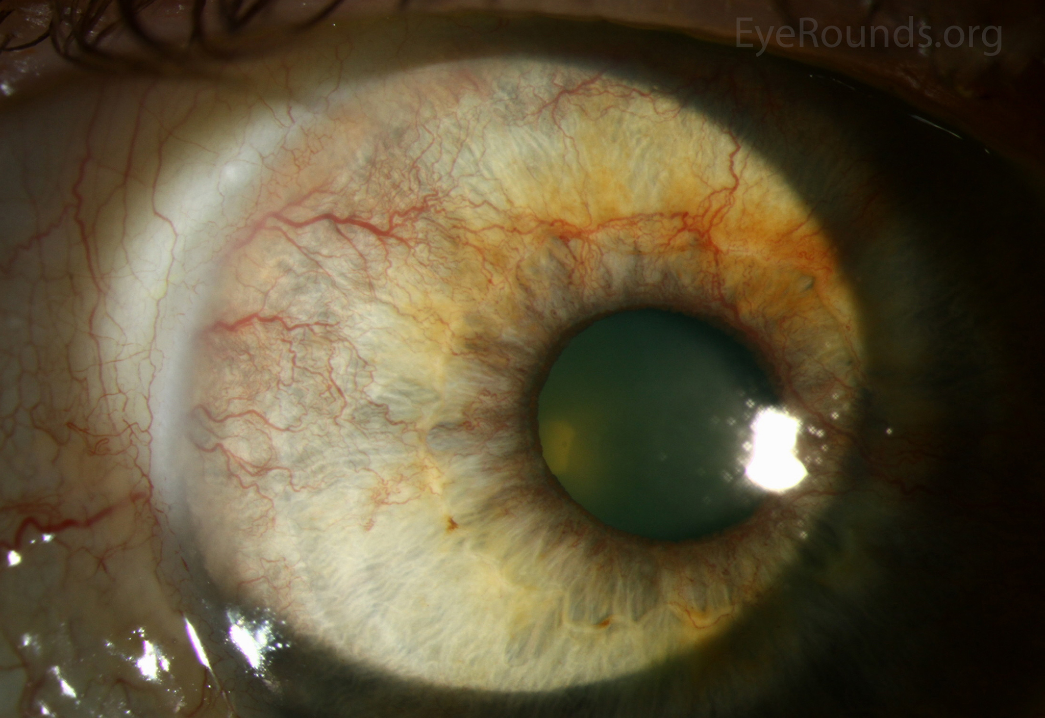 Image is from the Department of Ophthalmology and Visual Sciences, University of Iowa
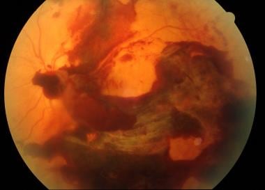
- Preretinal haemorrhages – bleed in the posterior hyaloid of the vitreous in front of the retina
- Show a fluid level because they are contained

- Vitreous haemorrhage – bleed within the vitreous humour due to permeable new blood vessels within the vitreous humour which can then bleed
- Can cause blindness
- Look like a blood haze in front of the retina
 (image from Medscape eMedicine )
- Retinal detachment– fibrosis can cause tractiondetachment
- Blindnessoccurs if the maculais involved
- This is the latest to develop of all complications
- Must be treated with a vitrectomy
- Nerve palsies due to ischaemia
- Optic neuropathy occurs due to ischaemia of CNII
- CNIII palsy
- Blindness occurs mainly due to macula involvement but also inretinal detachmentand vitreous haemorrhages
Investigations
There are several things to look at :
- Assess visual acuity
- Shows maculopathy
- E.g. using a Snellen chart
- Correct for best corrective vision therefore can differentiate between refractive error and pathology. I.e use glasses or pinhole
- Dilated eye exams
- Opthalmoscopy – less often used now
- Retinal photography – required for screening
- 2 photographs are taken
- Disc centred
- Macula centred
- Both are analysed and graded by the technician
- 2 photographs are taken
- Slit lamp biomicroscopy – used to assess patients as it can penetrate through cataracts unlike photography
- OCT scanning
- Shows topography of the retina and thus allows abnormalities to be picked up
- 7 layers of the retina
- Often used for the macula but can be used for the rest of the retina
- Used in patients who have maculopathy on photography
- Shows oedema well
- Can identify other pathological features:
- E.g. dot and blot haemorrhages occur in the middle layers
- Fluorescein angiography
- Finds leakages in detail
- Allows identification of neovascularisation
- Useful for planning laser photocoagulation
Management
Prevention
- Aim to control diabetes and risk factors to prevent onset and progression
- Hba1c control
- BP control – typically using an ACEI
- Cholesterol control – statin for 1º prevention
- Correct anaemia
- Ensure all diabetic patients are referred to retinal screening
- Annual ophthalmoscopy
Management
- See referral stages above (diabetic team => HES referral => urgent HES referral)
Conservative
- Optimise glycaemic control
- Eye aids could help with visual impairment patients
Medical
- Fenofibrate is also beginning to be used
- Was mainly an anticholesterol drug but there is evidence for preventative benefit of retinopathy
- Intravitreal Injections
- VEGF inhibitors (e.g. Leucentis, aflibercept, bevacizumab)
- Reduce neovascularisation
- Used in diabetic maculopathy but also central retinal vein occlusion and ARMD
- Steroid injections
- Can be used to reduce macular oedema
- Mainly used in patients who have already had cataract replacements and are not at risk of glaucoma
- VEGF inhibitors (e.g. Leucentis, aflibercept, bevacizumab)
Surgical
- Retinal photocoagulation
- Laser peripheral retina to reduce the ischaemic area and thus reduce the release of vasoproliferative growth factors that induce neovascularisation
- Used in R3/M1
- 3 strategies are used
- PRP is the main one for R3
- The other two strategies are used for Maculopathy
- Focal for smaller areas – focal maculopathy
- Grid treatment is used for diffuse maculopathy
- However it can relapse therefore follow-up is required
- Vitrectomy
- Used to clean out vitreal bleeds and replace vitreous gel with silicone to prevent neovascularisation