Summary
- Introduction to tracts
- The four ascending (sensory) tracts:
- Spinothalamic tract
- Dorsal column
- Spinocerebellar tract
- Trigeminothalamic tract

Introduction to Tracts
- Tracts are collections of the cell bodies of neurones that make up the white matter of the spinal cord, communicating information to/from the SC and brain
- Ascending tracts convey sensory information from SC to brain
- Descending tracts convey motor information from brain to SC

- Tracts receive the sensory axons from cells in the dorsal root ganglia (DRG)
- There are 4 main sensory tracts:
- Each is responsible for a different type of sensation
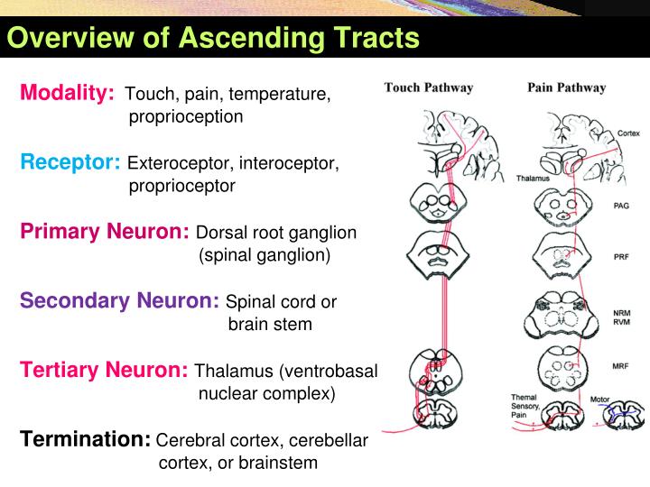
- Spinothalamic tract
- Perception of
- Pain
- Temperature
- Crude touch
- Firm pressure
- Includes two parts:
- Lateral spinothalamic tract
- Anterior/ventral spinothalamic tract
- Enters the spinal lemniscus in brainstem
- Perception of
- Dorsal columns
- Perception of discriminative (fine)touch
- Enters medial lemniscus in brainstem
- Spinocerebellar tract
- Perception of proprioception
- Goes to cerebellum
- Trigeminothalamic tract
- Takes all 4 senses from the face (CNV)
- Visceral afferents from CNV, CNVII, CNIX, CNX
Spinothalamic tract
- 2 parts with different functions:
- Lateral spinothalamic tract
- Pain
- Temperature
- Ventral spinothalamic tract
- Crude touch
- Firm pressure
- Lateral spinothalamic tract
- The lateral spinothalamic tract has ends in two places
- First terminates in the reticular formation of brainstem
- This induces reticular alerting response in the entire nervous system
- This therefore initiates a reflex resposne
- It then reports to the limbic system
- Indicates nature of the stimulus
- First terminates in the reticular formation of brainstem
-
Pathway:
- Primary neurone
- Cell body in DRG
- Synapses at ipsilateral dorsal horn
- May ascend/descend a little at Lissauers Tract
- Secondary neurone
- Decussates at the level it enters
- Lesion in SC will therefore cause contralateral loss of sensation
- The impulse travels in the ventral or lateral spinothalamic tract
- Synapses in the thalamus (in the ventral posterolateral nucleus)
- Decussates at the level it enters
- Tertiary neurone
- Starts in thalamus/VPL
- Travels in the spinothalamic lemniscus
- Synapses in the primary sensory cortex

Pathway of the spinothalamic tract.
- Primary neurone
Dorsal Column
- This is responsible for sensation of:
- Conscious proprioception
- Discriminative/fine touch

- 2 parts – both run in the dorsal column of white matter
- Both carry the same function – but from different levels
- Both are named according to the nucleus they synapse at
- Fasciculus Gracilis – Medial dorsal column
- Afferents come from lower limb (Sacral/lumbar)
- Synapses in Nucleus gracilis in medulla
- Fasciculus Cuneatus – Lateral dorsal column
- Fibres come from from upper limb (thoracic/cervical)
- Therefore present throughout spinal cord
- Synapses in Nucleus cuneatus in medulla
- Fibres come from from upper limb (thoracic/cervical)
- An aid to remember this is that ‘Major League Gaming needs LUC‘
- I.e. Medial, Lower limb, Gracilis => Lateral, Upper limb, Cuneatus
-
Pathway:
- Primary neurone
- Cell body lies in DRG
- Travels in either fasciculus up to medulla
- Synapses in the CNS at the medulla either at:
- Nucleus gracilus
- Nucleus cuneatus
- Secondary neurone
- Forms Internal Arcuate Fibres
- NB These decussate in medulla
- Synapses in the VPL in thalamus
- Travels to thalamus as the medial lemniscus
- Forms Internal Arcuate Fibres
- Tertiary neurone (same as spinothalamic tract)
- Starts in thalamus/VPL
- Synapses in the primary sensory cortex
- Primary neurone

Spinocerebellar Tract
- Communicates 2 sensations with the cerebellum:
- Unconscious proprioception
- Whole limb + postural movement
- 2 pathways:
- Ventral spinocerebellar pathway
- Dorsal spinocerebellar pathway
- Only 2 neurons in both pathways
- Primary neurone
- Identical for both pathways (unlike the secondary neurone)
- Cell body lies in DRG
- Synapses in the dorsal horn (Nucleus Dorsalis)
- Secondary neurone (Ventral SCP)
- Travel up spinal cord as Clarke’s column
- Decussate in spinal cord at level of entry
- Pass into cerebellum at midbrain via superior cerebellar peduncles
- Pass back into pons and decussate again into other cerebellum hemisphere via middle peduncles
- Travel up spinal cord as Clarke’s column
- Secondary neurone (Dorsal SCP)
- Travel up Clarke’s column
- Enter cerebellum at medulla via inferior cerebellar peduncle
- NB, does not decussate at all
- Primary neurone

Trigeminothalamic Tracts
- Input into the trigeminothalamic tract comes from several cranial nerves:
- CNV
- This receives general afferents from the skin of the face/head/neck
- Synapse in trigeminal ganglion
- CNVII, CNIX, CNX (visceral afferents from the rest of the body)
- Received via the spinal nucleus
- Synapse at parasympathetic nuclei in medulla
- CNV
- The Trigeminal nucleus has 3 parts with different afferent types in each part (NB this is different to the trigeminal ganglion)
- Mesencephalic nucleus (proprioception)
- Chief/primary nucleus (pressure, touch) – ventral trigeminothalamic pathway
- Essentially dorsal column for the face
- Spinal nucleus (pain, temp) – dorsal trigeminothalamic pathway
- Essentially spinothalamic tract for the face

The trigeminal nucleus complex. Note the difference between the trigeminal nucleus and trigeminal ganglion.
- Essentially spinothalamic tract for the face
-
Pathway:
- Primary neurone
- Cell body lies in trigeminal ganglion
- This is analogous to the DRG in the spine
- Except Mesencephalic nuclei (see below)
- Cell body in mesencephalic nucleus itself – not in trigeminal ganglion
- Synapses in the trigeminal nucleus in brainstem according to sensation (e.g. pain in the spinal nucleus)
- E.g. pain/temp in spinal nucleus
- Touch/pressure in chief nucleus
- Cell body lies in trigeminal ganglion
- Secondary neurone
- Decussates at level of entry into brainstem
- Travel up as trigeminal lemniscus
- Synapses in the VPM in thalamus
- Tertiary neurone
- Starts in VPM
- Passes through internal capsule
- Synapses in primary sensory cortex (to face division – more lateral than limbs)
- Primary neurone

Leave a Reply